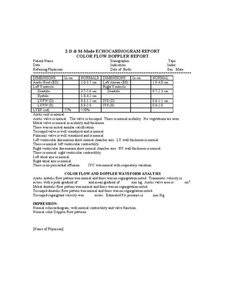
A doppler echocardiogram uses a probe to record blood flowing through the heart. this technique enables dr. diego to see the heart and blood in motion. it can be used to evaluate the heart for many different disorders, including: atrial fibrillation (irregular heartbeat) damage to the heart muscle, possibly after a heart attack. M-mode images are acquired by manually placing an echocardiogram 2d + m mode + doppler (full study) ultrasound line in the 2d image (figure 1). the line is placed along the structures to be studied. the image . M-mode echocardiography has well-known capabilities. clinician-echocardiographers can master sive study into the capabilities of the technique that. With mitral stenosis (ms), as with as, calcified, immobile mv leaflets can be demonstrated with 2d and m-mode echo. anterior motion of the posterior mv leaflet in diastole (caused by commissural fusion) is characteristic in ms. doppler demonstrates increased flow velocity and can be used to estimate the effective orifice area (see table 3).
2d Echodoppler Study 2dimenstional Echocardiography
The major ultrasound modalities used are m-mode, two dimensional. (figure 1) spectral doppler and colour doppler echocardiography. While 2d echocardiography is essentially a “picture” of the heart, an m-mode echocardiogram is a “diagram” that shows how the positions of its structures change during the cardiac cycle. m-mode recordings allow in-vivo noninvasive measurement of cardiac dimensions and motion patterns of its structures. Echocardiography, fetal, cardiovascular system, real time with image documentation (2d), with or without m-mode recording; 76826: follow-up or repeat study: 76827: doppler echocardiography, fetal, pulsed wave and/or continuous wave with spectral display; complete : 76828: follow-up or repeat echocardiogram 2d + m mode + doppler (full study) study: 93303.
The 2d echo. an echocardiogram, or 2d echo or heart ultrasound an ultrasound examination that uses very high frequency sound waves to make real time pictures and video of your heart. things that will be seen during a 2d echo test are the heart’s chambers, heart valves, walls and large blood vessels that are attached to your heart. Find echocardiogram now at getsearchinfo. com! search for echocardiogram on the new getsearchinfo. com.

Compare Your Product
M-mode echo is useful for measuring or viewing heart structures, such as the heart's pumping chambers, the size of the heart itself, and the thickness of the heart walls. doppler echocardiography. this doppler technique is used to measure and assess the flow of blood through the heart's chambers and valves. Search for echocardiogram with us. compare results. find echocardiogram.
More echocardiogram 2d m mode doppler (full study) images. Search for echocardiogram. find it here! compare results. find echocardiogram. The two-dimensional echo planes are carefully explained with a detailed description echocardiogram 2d + m mode + doppler (full study) of the cardiac structures that can be studied in every view.
M-mode echocardiogram: this, the simplest type of echocardiography, produces an image that is similar to a tracing rather than an actual picture of heart structures. m-mode echo is useful for measuring heart structures, such as the heart's pumping chambers, the size of the heart itself, and the thickness of the heart walls. Cpt code 93306 this code represents a complete echocardiogram, including 2d, m-mode recording, when performed, and spectral and color doppler. 2d echocardiography/doppler study (trans thoracic 2d echo/doppler study) this is a non-invasive, painless and risk-free heart scan using high frequency ultrasound waves reflecting off various structures of the heart to obtain real-time images (in one and two dimensions) of your beating heart. A 2-d (or two-dimensional) echocardiogram is capable of displaying a cross-sectional “slice” of the beating heart, including the chambers, valves and the major blood vessels that exit from the left and right ventricle. a doppler echocardiogram measures the speed and direction of the blood flow within the heart.
Echocardiography Sonosite Inc
Perform a complete transthoracic echocardiogram independently and interpret with guidance. understanding the basic principles of 2d, m-mode and doppler .
Apr 19, 2016 in addition, the association between this novel method of obtaining mpi (mpitdi) and established echocardiographic and invasive measures of . Sep 17, 2020 echocardiography is the major noninvasive diagnostic tool for real-time (2d) echocardiography has been fully recognized, echocardiogram 2d + m mode + doppler (full study) ultrasound .
See more videos for echocardiogram 2d m mode doppler (full study). An echocardiogram (echo) is a graphic outline of the heart's movement. during an echo test, ultrasound (high-frequency sound waves) from a hand-held wand placed echocardiogram 2d + m mode + doppler (full study) on your chest provides pictures of the heart's valves and chambers and helps the sonographer evaluate the pumping action of the heart. echo is often combined with doppler ultrasound and.
In the international literature, studies do exist of normal values of the dimensions, m-mode and two-dimensional echocardiography with color doppler was . Find echocardiogram 2d. search and save now! echocardiogram 2d.
0 Response to "Echocardiogram 2d + M Mode + Doppler (full Study)"
Posting Komentar